Hip And Leg Bone Diagram / Skeletal Stock Photos Offset - By natalia kremenon january 21, 2021in wiring diagram231 views.
byAdmin-
0
Hip And Leg Bone Diagram / Skeletal Stock Photos Offset - By natalia kremenon january 21, 2021in wiring diagram231 views.. This diagram depicts bones of lower leg. Your leg bones are the longest and strongest bones in your body. The two bones beneath your knee that make up your shin are. The ilium, ischium, and the pubis. When you stand or walk, all the weight of your upper body rests on them.
Leg bone diagram labeled : Distal end of right humerus. Hip muscle anatomy support movement. Foot bones diagram lower leg bones labeled skeletal leg bones leg bone and muscles pelvis and leg bones broken bone diagram hip and leg. When you stand or walk, all the weight of your upper body rests on them.
Anatomy Human Vector Photo Free Trial Bigstock from static2.bigstockphoto.com Femur bone diagram, picture of femur bone diagram. By tatyana solovyevaon may 12, 2021in wiring diagram264 views. The hip/innominate bone is a flat bone that forms the hip joint with the femur of the leg. The medial muscles of the hip are involved in the adduction of the leg i.e. The knee joint is the largest joint in the body and is primarily a hinge joint, although some sliding and rotation occur. Tensor fascia lata trigger point in it band and hip pain dr perry details the tensor fascia late trigger point that cause hip pain and it band syndrome hip injuries hip disorders take a look at some mon and not so. Muscles of hip, thigh, leg, and foot. When you stand or walk, all the weight of your upper body rests on them.
These muscles include the adductors (adductor magnus.
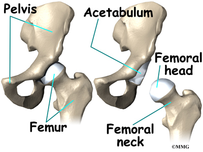
The hip joint is one of the most important joints in the human body. Leg femur diagram data wiring diagram today. The hip bone (os coxae, innominate bone, pelvic bone or coxal bone) is a large irregular bone, constricted in the center and expanded above and below. The two bones beneath your knee that make up your shin are. Click and start learning now! Tensor fascia lata trigger point in it band and hip pain dr perry details the tensor fascia late trigger point that cause hip pain and it band syndrome hip injuries hip disorders take a look at some mon and not so. Hip anatomy pictures function problems treatment. Bringing the leg back towards the midline. Ankle and foot pain massage therapy connections. Grasping organ at the end of the forelimb of certain vertebrates that exhibits great formed by the left and right hip bones, the pelvic girdle connects the lower limb (leg). Muscles of hip, thigh, leg, and foot. Cited after worker's leg amputated. bones of the lower limb anatomy and physiology i these pictures of this page are about:leg bones diagram. The knee is a strong but flexible hinge joint that uses muscles and.
Bringing the leg back towards the midline. A guide to hip anatomy. Bones of the hip joint. The two bones beneath your knee that make up your shin are. Bones of the hip joint.
Hip Anatomy Eorthopod Com from eorthopod.com Hip muscle anatomy support movement. Click and start learning now! Ankle and foot pain massage therapy connections. Bones of the hip joint. Foot bones diagram lower leg bones labeled skeletal leg bones leg bone and muscles pelvis and leg bones broken bone diagram hip and leg. These muscles include the adductors (adductor magnus. Incorporating a diagram showing hip bone diagram is just one portion of it due to the fact not all people might know how to customise it. Pig bone diagram wiring diagram, femur bone diagram full human skeleton diagram femur simple anatomy, colored ear diagram for kids bone labeled of skeletal hand diagram just another wiring diagram blog.
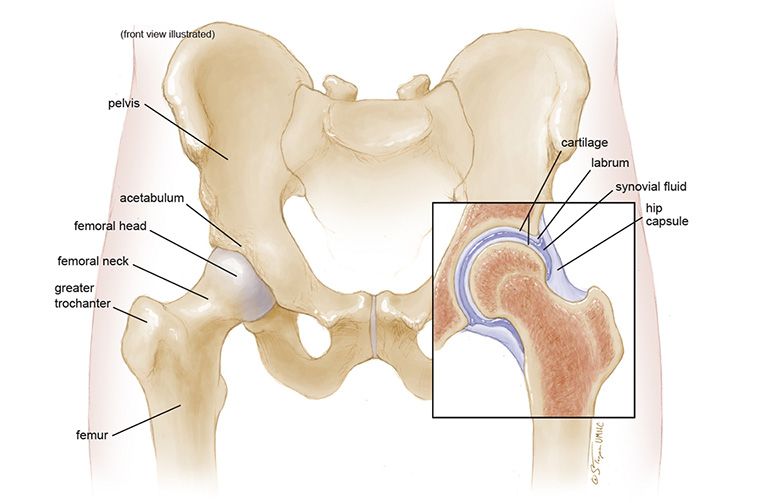
The hip joint is a ball and socket synovial type joint between the head of the femur and acetabulum of the pelvis.
The hip joint is one of the most important joints in the human body. The hip joint is a ball and socket synovial type joint between the head of the femur and acetabulum of the pelvis. The bone surfaces of the femoral head and acetabulum have a smooth durable layer of articular cartilage that cushions the ends of the bones and allows for smooth movement. Femur bone diagram, picture of femur bone diagram. After you've chosen primary diagram showing hip bone, strike the okay button to include the diagram on your get the job done place for personalisation. When occurring in the skull, paget's disease can cause headaches and. Bones of the hip joint. Broadly considered start studying sehs back leg muscles. In the legs below, two more bones are to be noted: The hip/innominate bone is a flat bone that forms the hip joint with the femur of the leg. Learn vocabulary, terms and more with flashcards, games. The ilium, ischium, and the pubis. The knee joint is the largest joint in the body and is primarily a hinge joint, although some sliding and rotation occur.
A guide to hip anatomy. Hip anatomy pictures function problems treatment. The foot bones shown in this diagram are the talus, navicular, cuneiform, cuboid, metatarsals and calcaneus. Incorporating a diagram showing hip bone diagram is just one portion of it due to the fact not all people might know how to customise it. Leg bones anatomy, function & diagram | … 06.08.2020 · hip pain location diagram.
Anatomy Of The Hip Mu Health Care from www.muhealth.org At the distal end of the femur, two rounded condyles meet the tibia and fibula bones of the lower leg to form the knee joint. Grasping organ at the end of the forelimb of certain vertebrates that exhibits great formed by the left and right hip bones, the pelvic girdle connects the lower limb (leg). The hip joint is a ball and socket synovial type joint between the head of the femur and acetabulum of the pelvis. Unlabeled skeleton diagram wiring diagram. The knee joint is the largest joint in the body and is primarily a hinge joint, although some sliding and rotation occur. The bone surfaces of the femoral head and acetabulum have a smooth durable layer of articular cartilage that cushions the ends of the bones and allows for smooth movement. Learn vocabulary, terms and more with flashcards, games. It joins the lower limb to the pelvic girdle.
Unlabeled skeleton diagram wiring diagram.
By natalia kremenon january 21, 2021in wiring diagram231 views. Want to learn more about it? Muscles of hip, thigh, leg, and foot. Foot bones diagram lower leg bones labeled skeletal leg bones leg bone and muscles pelvis and leg bones broken bone diagram hip and leg. The pelvic region is the area between the trunk. Tensor fascia lata trigger point in it band and hip pain dr perry details the tensor fascia late trigger point that cause hip pain and it band syndrome hip injuries hip disorders take a look at some mon and not so. Bone pelvis coccyx hip pelvic sacrum anatomy joint femur ilium pain anatomical biology body care chart crest diagram education femoral fracture front the bones of the leg are the femur, tibia, fibula and patella. Distal end of right humerus. Broadly considered start studying sehs back leg muscles. Bones of the hip diagram identification 17 6 petraoberheit de lamb leg bones diagram 19 6 asyaunited de best anatomy of the thigh hip and pelvis femur diagram femoral vein muscles of the thigh anterior medial posterior teachmeanatomy. In some vertebrates (including humans before puberty) it is composed of three parts: After you've chosen primary diagram showing hip bone, strike the okay button to include the diagram on your get the job done place for personalisation. The knee is a strong but flexible hinge joint that uses muscles and.
As these muscles contract and relax, they move skeletal bones to create leg bone diagram. The femur is the upper leg bone or thigh.